In order to properly understand how the
electron microscope works, it is necessary to explore some of the theories
and ideas that made the development of this device possible. In 1905 Albert
Einstein, in order to explain the photoelectric effect, postulated that light
was composed of quantized packets of energy which he called photons.
1 The energy of a photon is given by the following equation:
E = hc/8 (1)
where h is the universal constant known as Planck’s Constant. The numerical value of Planck’s Constant is:
h = 6.6260755 x 10⁻³⁴ Jqs.
This caused a great many people in the physics community no end of consternation because it had already been accepted that light was a wave since it could undergo interference and diffraction and only waves could exhibit such properties. Also, according to another one of Mr. Einstein’s great contributions to science, the special theory of relativity, every particle that has energy must also have momentum even if the particle has a rest mass of zero. The magnitude of the momentum of a photon is given by:
p = E/c (2)
where E is the energy of the photon, c is the speed of light, and p is the magnitude of the momentum of the photon.2 Combining the results of (1) and (2) gives the following interesting result:
p =
h/8 (3)
or
8 = h/p (4)
This result is interesting because it contains a relation between particle-properties (momentum) and wave-properties (wavelength).3
In 1924, Louis de Broglie proposed that perhaps all subatomic particles exhibited the same wave-particle duality that light exhibits.4 By relating the momentum of particles analogously to those of photons, he proposed that particles have a wavelength given by:
8= h/p = h/(mv) (5)
This wavelength is now known as the de Broglie Wavelength.5 De Broglie’s wave hypothesis received experimental verification in 1927.6 In 1927 Clinton Davisson and Lester Germer, while working at Bell Telephone Laboratories, were studying the surface of a piece of polycrystalline nickel by directing a beam of electrons at the surface and observing how many electrons bounced off at various angles.7 While conducting the experiment, air accidentally leaked into the vacuum chamber that the nickel was in and an oxide film was formed on the nickel.8 To remove this film, Davisson and Germer baked the specimen in a hot oven which, unknown to them, caused the nickel to anneal, creating large single-crystal regions on the specimen with crystal planes that were continuos over the width of the electron beam.9 When the experiment was repeated, the results differed greatly from their first observations.10 They found that strong maxima in the intensity of the reflected beam occurred at specific angles, in contrast to the smooth variation of intensity with angle that the two gentlemen had observed prior to the accident.11 Also, the angular positions of these maxima were dependent on the accelerating voltage, Va, used to produce the electron beam.12 Both gentlemen were aware of de Broglie’s hypothesis, and they noticed the similarity of their observations to x-ray diffraction.13 This led them to conclude that the electron beam in their experiment was being diffracted, thus verifying de Broglie’s hypothesis.14
It is also possible to express the de Broglie wavelength of a particle in terms of its kinetic energy. Consider an electron being accelerated from rest at point a to point b through a potential increase Vb - Va = Vba.15 The work done on the electron, eVba equals its kinetic energy, K.16 Using the fact that K = p2/(2m), we obtain the following:
eVba = p2/(2m), p = q(2meVba),
and the de Broglie wavelength of the electron is:
8= h/p = h/q(2meVba) (6)
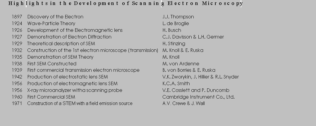
It is the above dual wave-particle nature of the electron that makes the scanning electron microscope, or SEM, possible. Not only do electrons possess wave properties, their wavelengths are much smaller than those of light (about .0054 nm depending on the accelerating potential).17 This would seem to indicate that a microscope which uses electrons instead of visible light to resolve an image would enable scientists to observe structures much smaller than those possible with an optical microscope. Click here to view a table that shows some of the important highlights in the development of Scanning Electron Microscopy18: