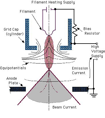
The scanning electron microscope differs from microscopes that utilize the transmission of light through a sample in order to resolve an image and transmission electron microscopes, which use the transmission of electrons through a sample to resolve an image. The electron beam is produced via an electron gun or filament. Click here for a diagram of the most common filament, the tungsten hairpin gun.
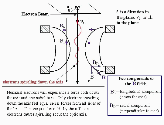
A voltage is applied to the tungsten filament (which acts like a cathode), causing it to heat up. When the filament gets hot enough, electrons are emitted thermionically. A strong electric force is present between the electrons emanating from the filament and the anode plate. This force causes the electrons to be accelerated towards the anode plate. In fact, some of the thermionic electrons are accelerated in such a manner as to stream right by the anode plate and down the column of the microscope to the sample. This electron beam is scanned back and forth over the surface of the specimen using electromagnetic lenses and the electrons that are reflected/emitted from the specimen are used to resolve an image on a cathode ray tube(CRT) or computer monitor. Click here for a diagram showing some of the forces experienced by the electrons as they pass through an electromagnetic lens.
Each magnetic lens consists of two cylindrical magnets that are either solid or made up of coils.
The optical column of the electron microscope must also typically be pumped down to a hard vacuum so that the filament does not oxidize (i.e. burn out) and to prevent air molecules from interfering with the electron beam. Electron microscope typically operate at a pressure low enough so that the mean distance that electrons in the beam travel before striking an air molecule is longer than the length of the microscope column.19 For most microscopes, the column is approximately 1 meter long and an air pressure of less than 1x10⁻⁴ mm Hg, or 1x10⁻⁴ torr, is needed and the normal working vacuum for SEMs with a tungsten filament is better than 5x10⁻⁵ mm Hg.20 Click here for a picture of a typical electron microscope illustrating the optical column.
This is how SEMs work at a very basic level. If this was all they did, they would be of very little interest to a physics student as an experimental tool. Fortunately, SEMs, unlike light microscopes, can analyze various interactions between the specimen and the electron beam to provide compositional data about the specimen.21 Some of the electron-specimen interactions that are observable are shown in Figure-4.
The primary specimen-beam interactions that physics and engineering students would be interested in are:
When the primary electron beam generated by the SEM strikes a specimen, the electrons in the beam will penetrate to a depth that is directly dependent on the energies of the electrons in the primary beam and inversely dependent on the atomic number(s) of the atoms in the specimen.22 Within the specimen, several scattering effects occur that are differentiable from one another by observing the relative energy lost by the beam electrons in their interactions with the atoms in the specimen.23 These interactions can be placed into two groups. In the first group, electrons are scattered due to elastic collisions with atoms in the specimen and lose very little energy in the process.24 These electrons are called backscattered electrons, typically have energies greater than 50 eV, and generally have energies of approximately 80% of the energy the beam electrons.25 In the second group, electrons collide with atoms in the specimen inelastically, causing the specimen atom with which it collided to ionize.26 Electrons emitted as a result of this ionization are called secondary electrons and generally are characterized as having energies less than 50 eV.27 These are the primary specimen-beam interactions, but other interactions are also observable. If a backscattered electron has enough energy to ionize other atoms in the sample before it escapes from the surface, more secondary electrons are produced.28 "Holes" left by the emitted secondary electrons must be filled with electrons from the outer shells and as the outer shell electrons move into inner shell "holes" energy must be discharged from the atom in the form of electromagnetic radiation (in the form of X-rays) or another electron with lower energy.29 Electrons emitted near the surface of the specimen in this sort of energy stabilization process is called an Auger electron.30 Auger electrons are usually most readily emitted by elements with a low atomic number (as opposed to X-rays which are usually emitted more readily from elements with a high atomic number) and can be used to determine the composition of the sample material.
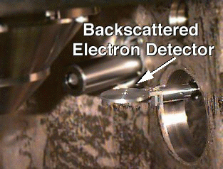
Backscattered electrons which have energies comparable to those of the incident beam behave in a fashion similar to beam electrons.31 Backscattered electrons travel in straight line paths that are impervious to the electrostatic attraction exerted by the low voltage secondary electron detector.32 The number of backscattered electrons produced in a beam-specimen interaction increases as the atomic number of the specimen atoms increases.33 Thus, if the atomic numbers of two (or more) groups of atoms in the specimen differ by more than three, these groups can be distinguished in the backscattered electron image.34 The element of higher atomic number will show up brighter on the screen(due to the fact that it produces more backscattered electrons).35 Due to the fact that backscattered electrons travel in straight lines, the detected backscattered electron signal is weaker than the secondary electron signal.36 The weakness of this signal can be demonstrated by the fact that it is possible to collect a strong secondary electron signal despite the noise contributed by the backscattered electrons because only the backscattered electrons directly in line with the detector will produce an image.37 In order to form an image with backscattered electrons, the secondary electron signal must be prevented from reaching the detector by removing the positive bias to the detector (which should be possible with the FSU Biology department’s SEM)38. Other method for obtaining a backscattered electron image are to use a microscope which has the ability to apply a slight negative bias to the detector, or to use modified scintillator detectors or other accessories.39 The area of the specimen from which the backscattered electrons are emitted is at least ten times larger then that of the secondary electron signal.40 If the beam current is increased for a stronger signal, the resolution of the backscattered electrons will differ from that of the secondary electrons by more than an order of magnitude.41 However, despite the lower spatial resolution, the backscattered mode is useful for atomic number contrast and topological contrast.42 Topological contrast makes use of the fact that backscattered electrons, since they travel in straight lines, may be obscured by a topological irregularity on the sample.43 This is useful in imaging relatively smooth specimens. Click here for a picture of a backscattered electron detector.
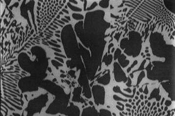
The following picture shows an example of using backscattered electrons to obtain compositional data:
This is an image of an aluminum-copper alloy formed using backscattered electron imaging. The light area is mostly aluminum and the dark area is mostly copper.
X-ray emission is one method of energy stabilization used by electrons that have been ionized by the incident electron beam. When an atom from an inner atomic shell has been liberated from the atom by the electron beam, an electron from an outer shell will fill the vacancy. When the electron moves from a higher energy state to a lower energy state, it may emit electromagnetic radiation in the form of X-rays. The x-rays emitted in this process have wavelengths that can be determined by the following equation:
Ehigh - Elow = hc/8 (7)
X-ray spectroscopy in the SEM involves the identification of a specific energy or wavelength for elemental analysis of the specimen.44 Then one of two types of detector are used: a wavelength dispersive spectrometer, which separates X-rays according to wavelength, or an energy dispersive spectrometer, which measures the energies of the X-rays.45 The relative peaks of the X-ray spectrum above the white-noise, Bremstralung radiation can then be used to determine the chemical composition of the sample.46
The SEM in the biology department at FSU currently only uses secondary electrons to form an image of the specimen. Secondary electrons are produced when the energy from the primary beam electrons causes the atoms of the specimen to ionize. As a result of this ionization, electrons are emitted from the sample. These electrons are called secondary electrons and have energies which are typically less than 50 ev.47 Due to their low energy (relative to the beam electrons) it is neccessary to "collect" the electrons with a positively biased collector in order to form an image. Bright areas on the image correspond to areas of the sample where large quantities of secondary electrons were emitted relative to other areas of the sample. Also, since the secondary electrons have low kinetic energies and are greatly attracted to the positively charged secondary electron detector, it is possible to form an image with secondary electrons that are slightly behind the specimen, relative to the primary electron beam. The result of this is that the SEM has a greater "depth of field" or viewable area than conventional optical microscopes.